
Veterinary Technicians’ Models Enhance Clinical Skills Education
Thanks to collaborative efforts between our professors and veterinary support staff, students have unique opportunities to learn clinical skills via a variety of delivery techniques, including specially made hands-on models.

Students engage with a model of a bovine prolapsed uterus, created by Animal Care Technologist Sara Gromley.
Thanks to collaborative efforts between our University of Arizona College of Veterinary Medicine faculty and veterinary technicians, our veterinary students have unique opportunities to learn clinical skills via a variety of delivery techniques, including specially made hands-on models. Through innovative models designed to enhance students’ understanding of vital techniques, CVM veterinary technicians like Chanda Hunt and Sara Gromley help facilitate student learning. We spoke with them about their insights into and role in the creation of these valuable learning tools.

Students learn to test for trichomoniasis using a model created by CVM's veterinary support staff.
Mirroring the collaborative nature of veterinary practice, our faculty and veterinary technicians work together to create models that enrich student learning. Our technicians, who draw from years of experience, work closely with professors who identify specific needs within the curriculum. These needs often manifest in the form of models that simulate various clinical scenarios, providing students with hands-on practice performing procedures crucial to their work in veterinary medicine.
Recently, our veterinary technicians have been busy creating models to aid students in learning various topics in large animal medicine.
Crafting Realistic Learning Experiences
When veterinary technicians are considering the best way to create a model, says Animal Care Technologist Chanda Hunt, they must keep in mind that it will be in iterative process. After being asked to create a model, the team creates a prototype that the faculty will evaluate and provide feedback for. Equipped with feedback and their personal experience of these processes and animals, the veterinary technicians will then either produce the model, or produce a different prototype.
One model Hunt created was designed to aid students in learning about male reproduction in large animals such as bulls and stallions. The model, featuring a box with a thin layer of nylon covering rubber silicone, allows students to practice carrying out rectal examinations, providing a unique opportunity to develop a vital skill. Hunt described the care she took in creating the model, sharing, “Dr. Eaton really wanted something with material behind the accessory gland, because in practice, you would feel something in between.” Veterinary support staff members’ attention to detail in simulating real-world scenarios is evident, ensuring that students gain a comprehensive understanding of the hands-on aspects involved in their future profession.
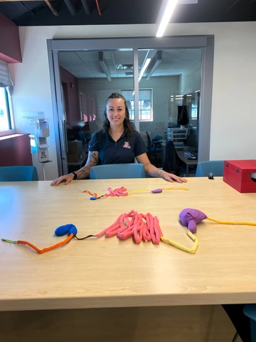
Chanda Hunt displays a portion of her model of a bovine gastrointestinal tract.
Students pair knowledge about bovine anatomy with a color-coded textile model of a bovine gastrointestinal tract, allows students to visualize the finer details of the GI tract, providing context to their theoretical knowledge. Hunt shared, “This model is a good visual, and I worked with the veterinarians to try to get the size right.” With feedback from the veterinarians, Hunt was able to craft an accurate model to aid students in their understanding of the animals they will work with.
Another model created by our veterinary technicians is the horse vulva Caslick model, also created by Hunt. Caslick operations can be vital to maintaining a female horse’s reproductive health, keeping the mare’s body from sucking air into the reproductive area. This model, created from silicone to stand up to multiple suturing attempts, is designed to enhance students’ surgical skills. Hunt explains, “This procedure is very important in broodmares and racing mares. If they have an open vulva, it should be closed using a Caslick operation.” This model goes beyond traditional techniques for teaching suturing, offering a more realistic experience by simulating the angle at which students would work during the procedure.
Visualizing Anatomy, Creatively
Sara Gromley, another of CVM’s Animal Care Technologists, is passionate about using her creative skills and veterinary know-how to impart knowledge to students through the model creation process.
One of Gromley’s designs, a bovine uterine prolapse model, allows students to engage with a process that might be daunting without preparation. Working with an actual-size model and learning what to do using the model in a lab setting aids students in developing comfort and proficiency with this technique.

This cleft palate model aids students in learning to assess the palate using only their sense of touch.
Among other recent designs by Gromley is her cleft palate model, created to aid students in identifying cleft palates in newborn foals. Gromley thought carefully about what would lead to the most productive learning experience and realized that it would be important to prevent students from seeing the palate models and diagnosing based on what they saw. This is not possible in real life, as it is not as easy to see into the animals’ mouths, so Gromley resolved to create models that mimicked this real-life limitation. Students rely on their sense of touch to identify foals with cleft palates, guided by their instructors. With the practice students gain in diagnosing and treating cleft palates, they become more prepared to support the health of newborn animals.
In addition to her work creating the GI and cleft palate models, Gromley also created a trichomoniasis model used for diagnosing a common venereal disease in cattle. She explained, “A bull will sexually transmit trich from cow to cow, which leads to fertility issues.” The model, designed to simulate the process of collecting smegma for diagnosis, enables students to practice the procedure without harming the animal.
Modeling Collaboration and Innovation
The importance of collaboration and communication between professors, veterinary support staff, and students cannot be underestimated. This joint effort ensures that the models not only meet educational goals but also provide an accurate representation of the clinical skills students will need to exercise in their future careers.

Sara Gromley noted how cooperative the work of creating and testing the models can be. She said, “I think it’s very rewarding. It’s wonderful because we all share expertise, and it’s very much a team effort.” The integration of diverse expertise—from the professors’ conceptualization to the technicians’ hands-on construction—supports a holistic learning experience for students.
Our CVM veterinary technicians expressed pride in their creations and were gratified to hear positive feedback from students. Chanda Hunt highlighted the impact of the models, saying, “In lab, the students have expressed that they find the models useful. The models help things click for them. They’re like, ‘Okay, I was reading it, and now I understand.’”
Our VetCats have incredible learning opportunities in part due to our veterinary support staff’s dedication to the accuracy, durability, and educational impact of the models they create. Through their experience and creativity, CVM’s talented veterinary support staff contributes significantly to providing an unparalleled clinical skills education and training day-one-ready veterinarians.